3D services

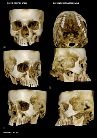
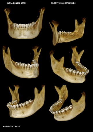
CBCT 3D imaging technology is far superior to conventional 2D dental imaging, as it enables practitioners to visualize all dental and facial structures in three dimensions and identify potential pathologic hard tissue lesions, provides superior and most accurate diagnosis and treatment planning in general dentistry, periodontics, implant planning, oral maxillo-facial surgical cases, and orthodontics.

IMPLANTS

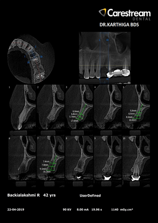
Measure quality and density of bone.
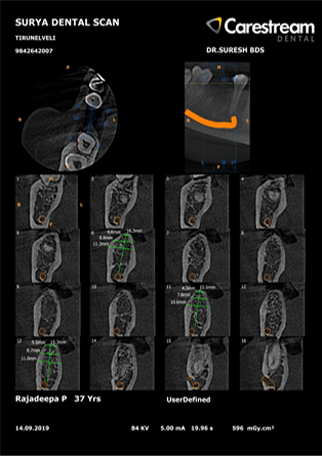
See and avoid critical anatomy.
Plan and determine proper placement of implant(s) while determining the most appropriate implant type and angulations.
1mm slices allows the Doctor to determine if cavitation is present before flap.
Determine whether bone grafting or sinus lifts may be necessary in areas where the amount of available bone is questionable.

ORAL SURGERY

Determine the precise three-dimensional position of a tooth within the alveolar bone and how this position relates to vital structures for extractions and impactions.
Plan and determine proper placement of implant(s) while determining the most appropriate implant type and angulations.
Trauma evaluation.
Visualize hard and soft tissues on the computer in three dimensions for planning maxillofacial surgeries.
Generate life-size CAD-CAM stereolithic (STL) models for surgical planning.

ORTHO ASSESSMENT

View bite in relation to bone structure for treatment plan.
3D evaluation of impacted tooth position and anatomy.
Orthognathic surgery treatment planning and growth assessments in true 1:1 imaging Planning for placement of dental implants for tooth restoration or orthodontic anchorage and for placement of temporary anchorage devices (TADs).
Assessment skeletal symmetry or asymmetry.

TMJ/TMD

Obtain a true 1:1 imaging of the condylar structures for more accurate assessments without superimposition and distortion.
3D evaluation of impacted tooth position and anatomy.
Orthognathic surgery treatment planning and growth assessments in true 1:1 imaging

PERIODONTISTS

Analyze periodontal bone defects on all sides of every tooth.
Assess the extent of furcation involvement.
Track the progression of advancing periodontal bone loss.
Treatment plan dental implants by evaluating bone parameters such as bone width, depth, and density.
Visualize vital structures such as the maxillary sinus, mental foramen and mandibular nerve prior to periodontal or implant surgeries.

ENDODONTISTS

More accurate identification and diagnosis of periapical endodontic pathosis than conventional radiography.
Visualization of obscure internal pulpal anatomy and root canal systems.
Assessment of internal and external root resorptive processes.
Identification of root fractures and other areas of trauma.
Volumetric and density comparisons of periradicular bone following endodontic treatment in order to assess the degree of success or failure Pre-surgical planning.

3D MOLAR/IMPACTIONS

Visualize an impacted tooth's position in relation to surrounding vital structures and nearby teeth and their roots.
Better assess the risk of treatment or non-treatment based on more accurate 3-dimensional analysis.

PATHOLOGY

CBCT scans provide a superior means of visualizing and studying pathological processes in the maxilla and mandible. This information is invaluable when planning any surgical efforts for biopsy or resection.
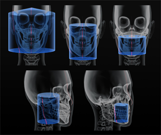
The data can be used to:
Render three-dimensional images of hard tissue abnormalities.
Provide more accurate information related to size, extent, location, and the relation to and effect on nearby anatomical structures.
Monitor the progression of the pathology as well as the success of treatment with the use of multiple scans.